Also known as: Venous doppler ultrasound
Duration: About 30 to 45 minutes
A duplex is a noninvasive test that combines both the doppler and the ultrasound. It can be performed on either your arteries or veins.
In a venous duplex, two different probes/ transducers (one for the doppler and one for the ultrasound) are used to measure how well the blood is flowing in the peripheral veins.

Uses:
- To check for any narrowings/stenosis in the veins of your legs
- To check how well the valves of the veins are working
- To check for blood flow – chronic venous insufficiency, varicose veins, deep venous thrombosis, and pulmonary embolism
- To check for blood clots
Preparing for the test:
![]() Download Pre Test Instructions
Download Pre Test Instructions
How it is performed:
- A technician applies a gel over the lower extremities (legs or ankles).
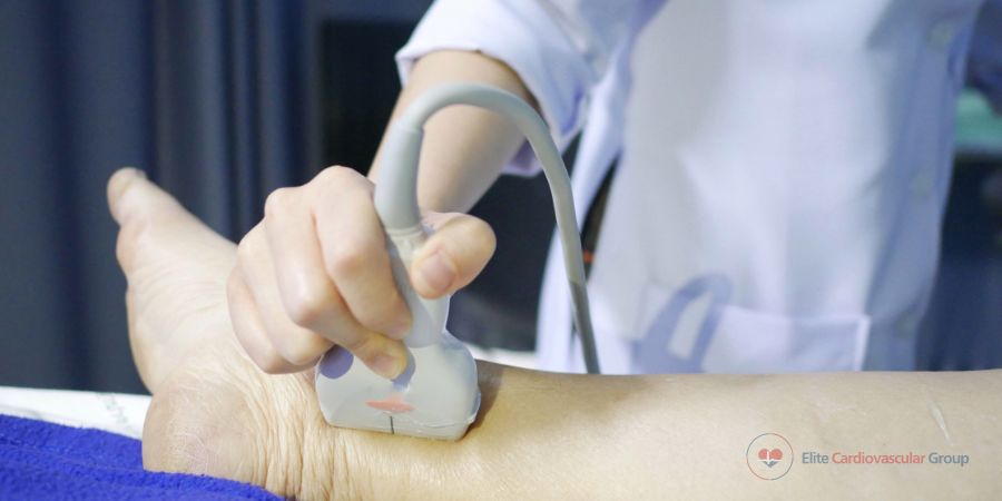
- The ultrasound probe is held firmly against your skin and glided in multiple directions as it sends out sound waves to the peripheral veins. Images are created when the probe picks up sound waves that bounce back from the veins.
- Then the doppler probe is held firmly against your skin and sends out sound waves to the red blood cells in the leg veins. The blood flow is heard and assessed for any stenosis/narrowings or blockages.
After the test:
- Your cardiologist will discuss the result with you.
- You can continue with your daily routine.
- Ask your cardiologist if you have any questions or concerns.
Show references
Baliyan V, Tajmir S, Hedgire SS, Ganguli S, Prabhakar AM. Lower extremity venous reflux. Cardiovasc Diagn Ther. 2016;6(6):533-543. doi:10.21037/cdt.2016.11.14
Necas M. Duplex ultrasound in the assessment of lower extremity venous insufficiency. Australas J Ultrasound Med. 2010;13(4):37-45. doi:10.1002/j.2205-0140.2010.tb00178.x
Khilnani NM, Min RJ. Imaging of venous insufficiency. Semin Intervent Radiol. 2005;22(3):178-184. doi:10.1055/s-2005-921950
